Maintaining healthy honey bees starts with vigilance. Early detection of threats can save a colony. Simple inspection may miss hidden problems that reduce productivity and survival.
Professional laboratory analysis gives a clear diagnosis when visual signs are unclear. Accurate samples must be collected and packaged to preserve results. Good sample handling speeds up answers and improves action plans.
Beekeepers should learn standard methods to gather samples and spot when experts are needed. Timely intervention protects the hive and limits losses across the apiary.
For additional resources and submission guidance, visit the bee health resources page.
Key Takeaways
- Early monitoring helps prevent colony collapse.
- One accurate sample leads to a reliable diagnosis.
- Proper collection and packaging matter greatly.
- Professional analysis uncovers hidden threats.
- Know when to seek expert help to protect your hive.
The Importance of Laboratory Testing for Honey Bee Diseases
Field observations can be misleading; precise analysis gives clear guidance. Visual checks may suggest a brood problem, but similar symptoms can signal different underlying issues.
Accurate diagnosis prevents unnecessary loss. Well-meaning beekeepers have sometimes removed frames or whole colonies based on a mistaken read of foulbrood signs. Confirming results avoids that risk.
Experienced bee inspectors and researchers stress that distinguishing American foulbrood (AFB) from European foulbrood (EFB) requires controlled analysis. This page links to a useful field diagnosis guide and explains when to request confirmation.
“Precise laboratory diagnosis ensures the right response and preserves long-term colony stability.”
| Sign | Common Cause | Needed Confirmation | Action |
|---|---|---|---|
| Spotty brood pattern | Multiple causes | Smear or culture | Send sample to lab |
| Sunken, perforated cells | Possible foulbrood | PCR or culture | Isolate, confirm |
| Unusual larval color | Bacterial or stress | Microscopy | Confirm then treat |
When in doubt, use professional services and practical tips such as how to sterilize hive tools. Confirming AFB, EFB, or other threats helps protect colonies and sustain production.
Identifying Common Symptoms in Your Apiary
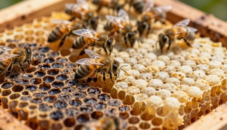
Small changes on the comb surface can signal major problems in the hive. Walk frames slowly and scan brood patterns. Healthy combs show a compact, nearly filled pattern with eggs, larvae, or pupae in most cells.
Brood pattern abnormalities often appear as a spotty or “pepperbox” brood pattern. Look for dark, concave, or punctured cappings. These signs frequently point to american foulbrood or european foulbrood, but they can also come from stress or poor nutrition.
Brood Pattern Abnormalities
- Spotty brood pattern is a primary indicator of foulbrood affecting larvae.
- Sunken or punctured cell cappings suggest a health crisis within the comb.
- Field observation is vital, but a formal diagnosis confirms the cause.

Adult Bee Behavioral Signs
Monitor adults at the entrance. Signs such as dysentery stains or bees unable to fly warn of wider colony stress.
“Act quickly on clear signs; early sampling and confirmation protect surrounding hives.”
| Sign | Likely Cause | Immediate Action |
|---|---|---|
| Spotty brood | Possible afb efb | Isolate frame; seek detailed disease guide |
| Sunken/punctured cell | Infection or stress | Remove suspect comb; document and sample |
| Adult dysentery / flightless | Systemic colony stress | Observe nearby hives; follow cleaning steps in cleaning plastic foundation |
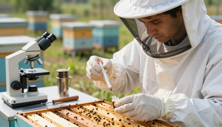
How to Collect Samples for Accurate Diagnosis
Good sampling depends on consistent technique and careful documentation. Take clear notes that list the hive number and apiary location. Proper labeling helps the lab link results to the right colony.
Collecting Larval Smears
Prepare one larval smear per suspect cell. Use a clean match or wooden stick to gently crush a larva onto a labeled glass slide. Spread into a thin smear and allow it to air-dry.
Seal the slide in a protective sleeve and record the sample ID. This simple step preserves material for fast laboratory diagnosis.
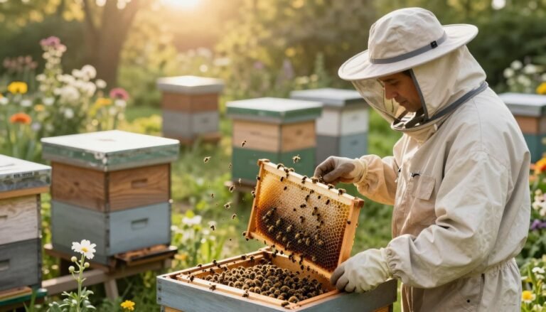
Harvesting Comb Samples
Cut a 5 x 10 cm piece of brood comb that contains the suspect larvae. Avoid any area with honey to prevent contamination.
- Wrap the comb piece in wax paper and then place it in a rigid container.
- Include hive number, apiary location, and date inside the package.
Use a strong cardboard box as a shipping device to prevent crushing during transit.
Sampling Adult Bees for Nosema
Collect about 30 sick or freshly dead bees from the ground near the entrance. Put them into a labeled vial and keep them cool.
Note that comb and adult samples answer different questions. If you need guidance on field methods like an alcohol wash, consult an expert guide such as the alcohol wash method.
“Clear labels, consistent technique, and secure packaging speed up accurate results.”
When ready to send material, follow submission steps on the site and submit a sample with all required information.
Understanding Laboratory Diagnostic Techniques
Modern diagnostic workflows separate spore-formers from transient microbes to protect colonies.
Preparation begins by heat shocking suspect material at 80 degrees Celsius for ten minutes. This step kills non-spore-forming bacteria while preserving spores for later observation.
After that, technicians inoculate BHIT agar and incubate plates at 34°C for 72 hours to let colonies develop. Cultural tests use specialized media because routine nutrient agar will not support these organisms.

Microscopic examination confirms the presence of spores and shows chain-forming bacterial morphology. Scientists at the USDA use these traits to tell american foulbrood apart from european foulbrood.
“Fluorescent antibody methods reveal specific pathogens as brightly fluorescing bodies under the scope.”
- Holst milk test detects proteolytic enzymes from suspect scale.
- Fluorescent antibodies offer rapid confirmation.
- Cultural and microscopic results together ensure an accurate diagnosis.
| Step | Purpose | Typical Result |
|---|---|---|
| Heat shock (80°C, 10 min) | Remove non-spore bacteria | Spore survival |
| BHIT agar (34°C, 72 hr) | Grow target colonies | Characteristic colonies |
| Microscopy / FA | Identify spores and antigens | Definitive confirmation |
For practical guidance on submission and further reading, see this bee diseases page.
Preparing and Shipping Samples to the Lab
A clear chain of custody starts with proper packaging and accurate labels. Pack each sample to protect fragile comb, frames, and adult specimens during transit. Good packing preserves evidence and speeds up results.
Packaging Requirements for Safe Transit
Follow strict steps to avoid contamination and damage. Wrap a comb piece in absorbent paper towel and place it in a sturdy cardboard box. Do not use plastic wrap or airtight containers, as condensation can promote mold and ruin tests.
- Send bees for Nosema analysis soaked in 70% alcohol and sealed in a leak-proof container.
- Label every sample with your registration number and collection date to speed processing at the site.
- When sending a piece of comb, include at least a 2×2 inch section with no honey so the diagnostician can find suspect larvae.
Always verify shipping rules with the referenced service. Post samples to Veterinary Sample Reception, Gribbles Veterinary Pathology, 1868 Dandenong Road, Clayton VIC 3168. For information on fees call 1300 307 190.
For guidance on sampler technique and field prep, see a practical sampler analysis guide and check the seasonal checklist at spring beekeeping checklist.
Conclusion
, Accurate field sampling and timely diagnosis give beekeepers the best chance to protect colonies. Short, consistent checks in the yard make problems easier to spot and document.
Follow the collection and shipping guidance on this page to identify a potential disease outbreak early. Use official laboratory services to secure correct results and the right treatment. Contact Customer Service Centre at 136 186 for specific information about submission and fees or visit the diagnostic labs and services page for more details.
A proactive beekeeper who pairs careful field observation with confirmation from qualified staff helps stop foulbrood and other threats. Quick action protects the honey bee and the wider apiary from a larger outbreak.