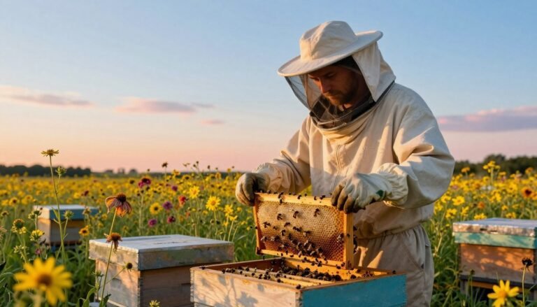
Detecting early threats keeps a hive healthy. Beekeepers must tell the difference between harmless spoilage in stored food and infections that harm larvae.
American Foulbrood, caused by Paenibacillus larvae, can destroy an apiary because its spores persist on equipment and comb for years. European Foulbrood and chalkbrood also affect larvae but come from different pathogens.
Routine inspections focus on cells and larvae, queen strength, and worker behavior. Strong colonies with active worker bees resist wax moths and other scavengers better than weak colonies.
Good sanitation of equipment and careful handling of comb and honey lower the risk of spores spreading. For detailed disease ID and treatment, consult a reliable guide on honey bee diseases and pests, and for moisture and mold control see tips on preventing mold inside a beehive.
Key Takeaways
- Learn to spot early signs in cells to protect larvae and adult bees.
- American Foulbrood spores are persistent; equipment hygiene is critical.
- Strong queens and robust worker populations are the best defense.
- Keep comb dry and ventilated to reduce fungal growth and spoilage.
- Use clean tools and protocols during inspections to limit spread.
Understanding the Hive Environment
A hive’s microclimate and layout shape how bees build comb and rear larvae.
Airflow, elevation, and dry frames lower fungal risk and keep stored honey and food in good condition. After winter, many colonies contain older workers and need extra care to protect the brood and support the queen.
Infected nurse bees can pass viruses to larvae through brood food, so clean, active workers matter. Wax moth larvae prefer dark comb rich in entrapped pollen and larval skins; the greater wax moth is a natural scavenger of old comb.
“A well-ventilated hive, elevated above damp ground, helps prevent mold and supports strong spring buildup.”
- Manage moisture to protect cells and stored reserves.
- Monitor after winter so nurse bees can feed healthy larvae.
- Keep frames dry to deter wax moths and fungal growth.
- Learn how to prevent chalkbrood as part of environment control.
Distinguishing Moldy Pollen vs Diseased Brood
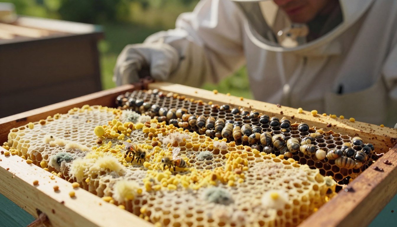
Beekeepers must use clear visual checks to tell if a hive problem comes from spoiled stored food or an infection harming larvae.
Visual Cues
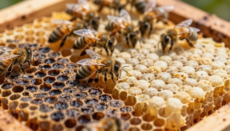
Inspect individual cells and note color and texture. Infected larvae may turn off-white, yellow, brown, or gray. You might see tracheal tubes beneath the skin in severe cases.
Dead larvae can form a rubbery scale that workers sometimes remove, but heavy infections overwhelm the colony’s cleanup. The larval head capsule often darkens to brown as the condition progresses.

Environmental Factors
High humidity and poor ventilation let common molds grow on stored comb after a hive dies during winter. This looks different from sunken or perforated cappings seen with many larval diseases.
Queen performance also matters: a patchy brood pattern points to colony stress, while spoiled food more often signals inadequate airflow or moisture control.
- Observe color and texture of larvae across several frames.
- Note capping shape: sunken or punctured cappings suggest disease; fuzzy or powdery growth suggests storage spoilage.
- Check ventilation and honey stores when winter losses occur.
| Feature | Storage Spoilage | Larval Infection |
|---|---|---|
| Common look | Fuzzy or slimy patches on comb | Off-white to gray larvae; head capsule browning |
| Cappings | Usually intact; may show mold near food | Sunken, perforated, or discolored cappings |
| Colony signs | Dead or absent colony after winter; low activity | Workers removing scales; poor brood pattern |
| Environmental driver | High humidity, poor ventilation | Pathogen presence; stressed or young nurse bees |
When in doubt, document findings and consult lab guides. For detailed diagnostic resources see the USDA honey bee disease guide and read about related cold-incubation signs at identifying chilled brood.
Identifying American Foulbrood
A rapid field test helps determine whether a spotty brood pattern comes from a lethal infection.
American Foulbrood is caused by Paenibacillus larvae and can devastate an apiary because spores survive on equipment and comb for decades.
The Ropiness Test
The ropiness test is a simple on-site check. Insert a clean twig or match into a suspicious cell and gently pull out the remains.
If the contents stretch up to 3/4″ before snapping back, the result strongly suggests AFB. Photo examples by Jon Zawislak show this clearly in practice.
“If the mixture strings out up to 3/4” before snapping back into the cell, it is very likely infected with AFB.”
Action steps:
- Do not move frames between colonies; spores spread on tools and adult bees.
- Contact your state apiary inspector if you suspect infection; many states require containment by burning infected hives.
- Never reuse old equipment without proper lab testing or approved sterilization; spores resist heat and cold.
For diagnostic details and lab guidance, consult the UADA beebrood conditions resource and learn about safe equipment repairs at beebrood conditions and repairing damaged hive boxes.
Recognizing European Foulbrood
Watch the open brood nest closely in spring: European Foulbrood, caused by Melissococcus plutonius, kills young larvae before their cells are capped. Inspect multiple frames for off-white, yellow, brown, or gray larvae and look for twisted or discolored remains.
A spotty brood pattern and poor brood distribution signal a problem. Nurse bees can contaminate brood food and spread the infection when weather and the colony population are weak after winter.

Intervention often works without antibiotics. The shook swarm technique lets beekeepers move adult bees onto clean comb and fresh foundation to break the infection cycle. Removing the queen for about two weeks can also help the hive rebuild healthy brood and resources.
“Early detection and prompt management give most colonies a good chance to recover.”
- Check open cells across the nest for discolored larvae and an uneven pattern.
- Use shook swarm and brood breaks before resorting to chemical options; EFB does not form persistent spores like other threats.
- For guidance on when to remove or discard old comb after an infection, see when to discard comb after disease.
Managing Sacbrood Virus
Sacbrood virus affects bees worldwide and shows up when capped cells hold larvae that fail to pupate.
Infected larvae sit on their backs in a distinct “canoe” posture. When you pull one from the cell it can appear as a bag of liquid.
There is no practical medical cure. Beekeepers must use management steps to break the cycle and protect the hive.
“Caging the queen for two weeks often halts transmission to young bees and gives the colony time to recover.”
Practical steps include caging the queen for about 2 weeks and ensuring the colony has extra food and comb resources during spring recovery.
- Monitor brood patterns and nurse bee strength.
- Consider requeening if poor performance continues; nurse bees pass the virus in brood food.
- Limit movement of comb and equipment between colonies to reduce spread.
| Issue | What to Watch | Action |
|---|---|---|
| Larval sign | Canoe posture; liquid when removed | Document, isolate frame |
| Colony impact | Poor brood pattern in spring | Cage queen 2 weeks; add resources |
| Long term | Recurrent infection | Requeen and improve nurse bee nutrition |
For guidance on reporting and inspection, contact your inspector or see the detailed note on sacbrood virus reporting.
Dealing with Chalkbrood and Stonebrood
Fungal infections such as chalkbrood and stonebrood appear as hardened larval remains that tell a different story than bacterial threats. These fungi attack larvae after they ingest spores, and signs often show where worker bees cannot fully remove the remains.

Chalkbrood Mummies
Chalkbrood is caused by Ascosphaera apis. Infected larvae become mummified and often show white to gray, brittle bodies. You may spot these mummies on the landing board or among cells.
A strong colony population helps remove mummies. Still, beekeepers must scrape and clean woodenware and replace old brood comb to stop spread.
Stonebrood Risks
Stonebrood stems from Aspergillus species and is rarer but serious. Mummies can be yellow, green, or black and grow very hard.
“These fungal spores can pose a respiratory risk to people handling heavily infected equipment.”
Wear protection when scraping and sterilize tools. If the condition persists, replace comb, elevate the hive to reduce water, and improve ventilation.
- Monitor for 2–3 days in spring to see if the infection clears with warming weather.
- Clean and sterilize equipment; discard heavily infected frames.
- Support the queen and worker bees so the colony can remove mummies and rebuild strong brood patterns.
| Feature | Chalkbrood | Stonebrood |
|---|---|---|
| Pathogen | Ascosphaera apis (fungus) | Aspergillus spp. (fungus) |
| Signs | White/gray brittle mummies on landing board or cells | Yellow/green/black hard mummies; rare |
| Risks | Colony cleanup burden; weaker brood pattern | Human respiratory risk when handling spores |
| Management | Clean woodenware, replace comb, elevate hive | Sterilize tools, replace frames, use PPE when cleaning |
Assessing Black Queen Cell Virus
Black Queen Cell Virus (BQCV) kills developing queens after their cells are sealed. The wax often looks dark and oily when infection advances.
BQCV is found worldwide and can remain latent in healthy colonies. Incidence usually peaks in early spring when many queen cells are made.
The virus spreads mainly through contaminated brood food. While varroa have carried BQCV, they are not the primary vector for queen infection.
“Watch queen rearing operations closely; simultaneous cell production raises risk.”
Practical checks:
- Inspect queen cells for dark, sunken cappings and oily resin.
- Limit large-scale queen rearing in a single hive to reduce spread.
- Manage stressors like nosema to help the colony handle latent infections.
| Feature | What to look for | Recommended action |
|---|---|---|
| Appearance | Dark, oily sealed cells; dead larvae inside | Remove affected cells; isolate queen rearing area |
| Timing | Peaks in spring during queen production | Stagger rearing; monitor nearby colonies |
| Vector risk | Varroa found with virus but not main vector | Control varroa anyway to support development |
Evaluating Secondary Symptoms like Bald Brood
Straight rows of open cells can be a clear sign of pest activity rather than a primary infection. Bald brood is a symptom caused when small caterpillars tunnel through comb and force worker bees to uncap brood cells to remove intruders.

Wax Moth Infestations
Galleria mellonella larvae bore through wax and leave tidy lines of uncapped cells. Workers try to catch the caterpillars, so the pattern often runs in straight rows for several cells.
As noted in photos by Ashley Mortensen, these uncapped rows show active cleanup. Bald patches are not a disease, but they reveal that the hive may be weakened.
Prevention focuses on strength. Keep strong colonies that guard the entrance and repair damage. Seal hive bodies well to block moth entry and reduce comb destruction.
| Feature | Cause | Action |
|---|---|---|
| Bald brood pattern | Wax moth larvae tunneling | Strengthen colony; seal hive; remove damaged comb |
| Visible uncapped rows | Worker bees removing caterpillars | Inspect frames; support worker bees; clean equipment |
| Long-term risk | Weakened hives; increased varroa pressure | Monitor for mites; requeen or combine if weak |
Addressing Parasitic Mite Syndrome
Parasitic Mite Syndrome (PMS) results when heavy varroa infestations pair with high viral loads. The result is a weakened hive where spotted brood patterns and dead larvae appear in cells.

Photos by Michael E. Wilson (beeinformed.org) show how colonies with PMS display scattered sealed cells and adult bees that cannot maintain the nest. You may see bees with deformed wings crawling near the entrance or on the ground.
Immediate action matters: treat mites with an effective, registered varroa control. Provide supplemental food and water if the colony is weak, and consider consolidating frames into a smaller hive to help worker bees feed the queen and larvae.
- Break the brood cycle for a few days to reduce mite reproduction and viral spread.
- Use approved mite treatments and follow label directions for safety and effect.
- Monitor honey stores and replace or remove heavily infected frames; spores and mites travel on wax and equipment.
“Left untreated, PMS leads to total colony collapse as worker bees fail to support the hive.”
| Sign | Action |
|---|---|
| Spotty brood pattern / dead larvae in cells | Treat varroa; consider brood break for several days |
| Deformed wing adult bees at entrance | Isolate frames; check mite loads; provide food and water |
| Rapid population decline | Consolidate hive; seek advice from beekeepers or inspector |
Conclusion
Consistent hive checks let you spot abnormal patterns and act while recovery is possible.
Recognizing the signs of moldy storage and problems in developing larvae is the first step to a healthy apiary. Learn the specific causes and how environment, nutrition, and handling affect outcomes.
Keep a schedule of inspections and record findings. Early attention to any disease or infection saves frames, time, and expense. Follow local rules and contact your state apiary inspector for suspected American Foulbrood.
A well-managed hive with dry comb, good ventilation, and strong workers is your best defense against seasonal threats.
FAQ
What are the key visual differences between moldy pollen and diseased brood?
Healthy stores of pollen appear granular and brightly colored. Spoiled stores often look discolored and may show fuzzy growth or an off odor near capped cells. In contrast, infected larvae or pupae show abnormal color, sunken cappings, or irregular patterns in brood comb. Look for dead larvae that are twisted or liquefied, and worker behavior changes around affected cells. Check for Varroa mites on adults too, since mites often accompany brood disease.
How does hive environment influence these problems?
Cold, damp, or poorly ventilated hives increase humidity and favor fungal or mold growth in stored food. Overcrowding, limited forage, and weak colonies stress bees and make brood more vulnerable to bacterial and viral infections. Regular inspection, good ventilation, and ensuring adequate food and water reduce risk. Beekeepers should also monitor queen health and worker population trends.
What visual cues help distinguish contaminated stores from infected larvae?
Contaminated stores often sit in peripheral cells and show surface growth without internal collapse. Infected larvae display degradation inside brood cells: discoloration, sunken caps, or a ropy, sticky consistency for some bacterial diseases. Note patterns—scattered spoiled stores suggest storage issues, while clustered affected brood points to infectious spread.
Which environmental factors most promote brood infections?
High humidity, prolonged cold, and poor airflow favor fungal pathogens. Stressors such as nutritional deficiency, queen failure, and heavy Varroa mite loads weaken colony immunity and let bacteria or viruses spread. Seasonal weather swings in spring or fall often trigger symptom outbreaks.
How do beekeepers identify American Foulbrood (AFB)?
AFB causes a distinctive sticky, dark residue when infected larvae decompose. The “ropiness” of larval remains is a key sign. AFB spores are highly resilient and spread via equipment, bees, and beekeeper activities. Confirmatory lab tests are recommended and many states require reporting and specific control measures, such as sterilization or destruction of infected equipment.
What is the ropiness test and how is it performed?
The ropiness test involves probing a suspicious capped cell with a sterile stick. If decayed larval material stretches into a ropy string, that supports an AFB diagnosis. Exercise caution: use new tools, isolate samples, and follow local regulations. Positive results usually prompt immediate containment and lab confirmation.
How can I recognize European Foulbrood (EFB) in my hive?
EFB often shows uneven brood patterns, twisted or discolored larvae, and sunken or perforated cappings. Unlike AFB, larvae may not show the classic ropy pull. EFB typically affects younger larvae and is associated with colony stress, poor nutrition, or cooler temperatures. Treatment and management focus on boosting colony strength and, when needed, approved antibiotic therapy following regulatory guidance.
What are common signs of Sacbrood Virus?
Sacbrood causes dead larvae to dry into a sac-like appearance with a distinct yellow or brown tint. Affected cells often hold mummified larvae that lie on their backs with a pointed head. The virus spreads via nurse bees and contaminated comb. Managing colony stress and requeening can help reduce impact.
How do I spot chalkbrood and stonebrood, and what actions help?
Chalkbrood produces white or gray mummies that look like chalk and often protrude from cells. Stonebrood, caused by Aspergillus species, forms dark, hard mummies and can infect bees and equipment. Good airflow, avoiding excess moisture, replacing old comb, and maintaining overall colony health reduce fungal risks. Severe stonebrood may require apiary sanitation and professional advice.
What do chalkbrood mummies look like and where are they found?
Chalkbrood mummies are brittle, pale, and may fall to the bottom board or lie in cells. You’ll find them near brood frames in damp or spring conditions. Their presence signals fungal activity and often correlates with weak or stressed colonies. Improve ventilation and colony vigor to prevent recurrence.
What risk does stonebrood pose to colonies and beekeepers?
Stonebrood can kill larvae and produce dark, calcified mummies. Aspergillus spores can be abundant in contaminated equipment and pose a risk to colony health. Decontaminate or replace highly affected comb and maintain dry, well-ventilated hives to limit spread.
How can I detect Black Queen Cell Virus in my apiary?
Black Queen Cell Virus causes queen pupae and cells to turn dark and die, often producing an irregular brood pattern. Look for shriveled or blackened queen cells and sudden queen failure or poor colony development. Vectoring by Varroa mites and other stressors increases incidence. Good mite control and requeening with robust stock help reduce risk.
What secondary symptoms, like bald brood, should alarm me?
Bald brood—naked larvae exposed by missing cappings—indicates nurse bee problems, nutritional deficiency, or viral infections often linked to mite damage. Check adult bee population, food stores, and signs of Varroa mite infestation. Address underlying causes rather than only treating symptoms.
How do wax moth infestations appear and how can they be prevented?
Wax moths leave webbing, tunnels, and shredded comb, often in weak or stored colonies. Infested frames show cocoon fragments and irregular comb damage. Prevention includes maintaining strong colonies, freezing or heat-treating stored comb, and using sealed storage. Regular inspections and prompt removal of affected equipment limit spread.
What is Parasitic Mite Syndrome (PMS) and how does it relate to brood diseases?
Parasitic Mite Syndrome describes colony collapse symptoms caused by heavy Varroa mite infestations and associated viruses. Signs include spotty brood patterns, deformed adults, and reduced population. Mites vector viruses that weaken brood development and can precipitate other bacterial or fungal problems. Implement an integrated pest management plan combining monitoring, timely treatments, and good husbandry to protect your colony.