Reviewed by Thomas B. · How we review
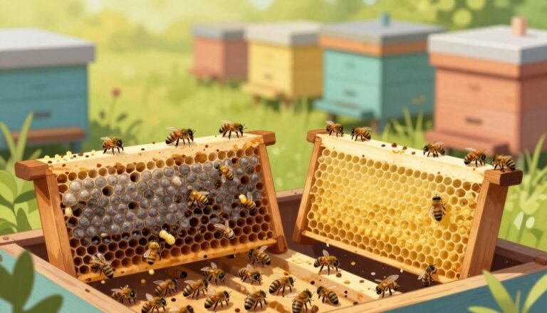
Early inspection and good equipment care are the backbone of healthy hives. Spring and fall checks help beekeepers spot odd cell caps or off‑colour larvae before problems spread. Quick, regular reviews protect colonies and the larger apiary.
Recognizing unusual signs — sunken or misshapen cappings, scattered patches across central frames, or larvae that sit oddly in cells — should prompt further action. Those clues can signal disease or stress rather than simple temperature loss.
Novice beekeepers should study safety and sampling guides before opening hives. Professional keepers must remain vigilant: early detection preserves entire colonies and limits pathogen spread through shared equipment.
For guidance on foulbrood symptoms and testing options see resources on foulbrood identification and practical notes on chilled brood assessment. Sanitation, proper storage, and timely equipment rotation reduce risk across hives.
Key Takeaways
- Inspect frames in early spring and autumn to catch problems early.
- Watch for unusual cappings, off‑colour larvae, and abnormal positions.
- Maintain strict equipment hygiene to limit pathogen spread.
- Train inexperienced handlers on safety and sampling before inspections.
- Use official resources and testing for accurate diagnosis and action.
Table of Contents
- Understanding Larval Discoloration in Bee Brood
- Identifying Healthy Brood Patterns
- Common Bacterial and Fungal Causes of Discoloration
- Diagnostic Techniques for Hive Inspections
- Managing and Controlling Infected Colonies
- Best Practices for Apiary Sanitation
- Conclusion
- FAQ
Understanding Larval Discoloration in Bee Brood
A clear grasp of how larvae change inside cells makes diagnosis simpler.
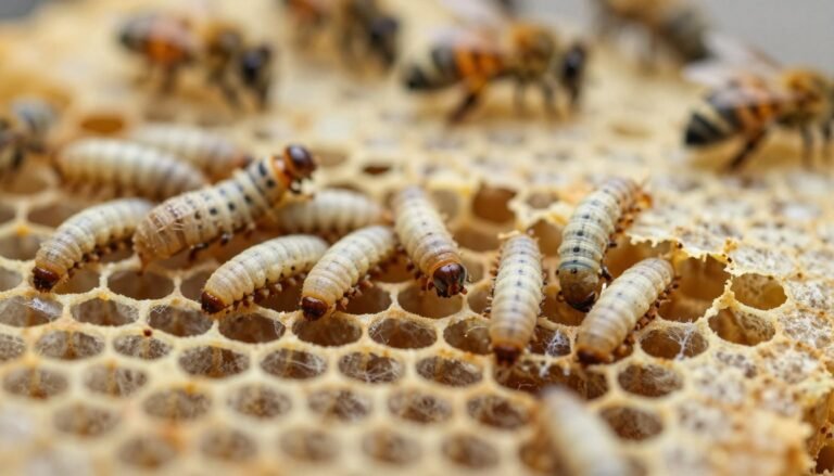
Brood includes eggs, larvae, and pupae that occupy the comb cells in the broodnest. Young larvae are curled, pale grubs that sit like a small letter ‘C’ at the base of a cell before they grow to fill it.
The pupae stage follows feeding. During pupation the larva transforms into an adult while sealed in its cell. Watching these stages helps observers tell normal growth from trouble.
When patterns become patchy or cells show odd contents, the colony may have a disease. Irregular spacing of healthy individuals is one of the earliest signs to note during an inspection.
Quick checks of development help you decide next steps: sample, test, or treat. For practical diagnostic steps see the field diagnosis guide.
| Stage | Appearance | What to watch for |
|---|---|---|
| Egg | Tiny, upright filament | Present in every healthy cell |
| Larva | Curled white grub (C-shape) | Uniform size; no sunken cappings |
| Pupa | Immobile, changing form | Sealed cell; regular timing to emergence |
| Warning signs | Patchy pattern or odd cell contents | Investigate; see prevention tips like chalkbrood prevention |
Identifying Healthy Brood Patterns
Good brood coverage and clear cell contents are the first things to check when you inspect frames. A steady, even patch of developing brood across a frame shows a well‑mated queen and attentive workers.
Visual Characteristics of Healthy Larvae
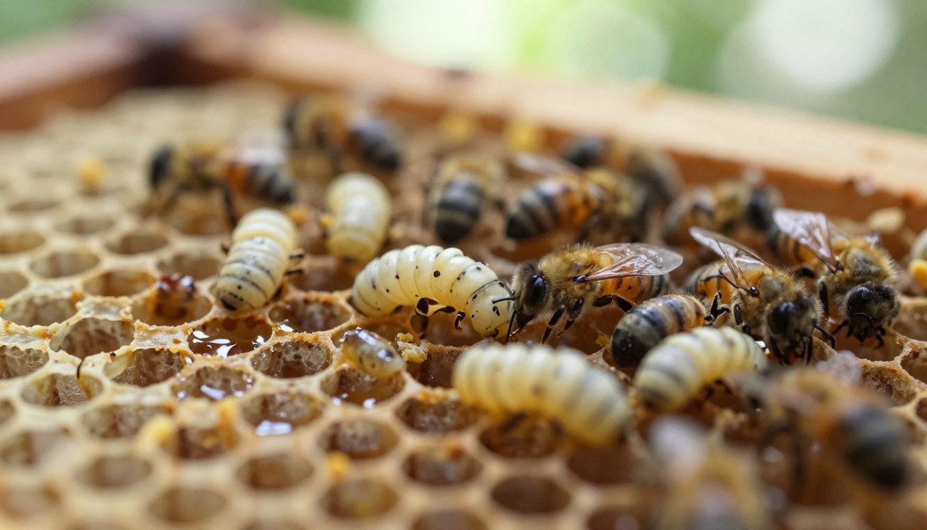
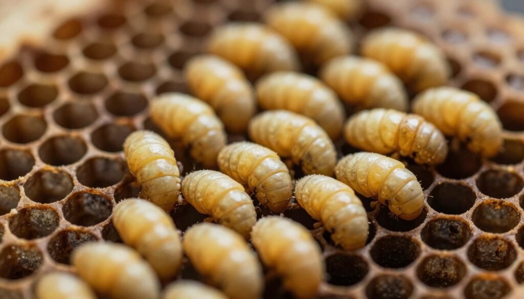
Healthy larvae are glistening and pearly white. You can often see an orange gut line along the back of each larva as it grows in the cell.
Look for nearly every nearby cell to hold a developing individual. Patches with many empty spots may point to laying or feeding issues.
Healthy pupae start white and darken as adult features appear, first in the eyes of the developing worker.
Recognizing Normal Capping Appearance
Brood caps are usually cream, light brown, or tan and should be uniformly convex.
Caps must be intact without holes or perforations. Sunlight tilted across the frame helps reveal subtle irregularities—hold the top bar near your stomach and tilt the frame to the light.
| Feature | Normal | Note |
|---|---|---|
| Cap color | Cream to tan | Uniform convex shape |
| Cell fill | Most cells occupied | Compact pattern signals strength |
| Pupae | White to darkening | Eyes darken first |
For more on managing hive pests and signs to watch, see common hive pests.
Common Bacterial and Fungal Causes of Discoloration
Several bacterial and fungal agents produce telltale signs that experienced keepers learn to spot quickly. Recognizing distinct patterns in cells and remains speeds diagnosis and guides control choices.
American Foulbrood Symptoms
American foulbrood is caused by Paenibacillus larvae, a spore‑forming bacterium that devastates a colony. Infected cells may contain sticky, ropy remains that can be stretched 25mm or more with a matchstick.
Frames with this disease often require strict removal or destruction and careful handling of equipment to prevent spread.
European Foulbrood Characteristics
European foulbrood typically affects curled larvae while still in the stage. The remains have a soft, watery feel that soon turns pasty and discolored.
Unlike the ropy test of American foulbrood, these cases show rapid change in consistency and scattered patchy comb patterns.
Fungal and Viral Infections
Fungal infections such as chalkbrood kill larvae about two days after capping; dead pupae harden into white, chalk‑like mummies.
Sacbrood virus produces a sac‑like larva with the head lifted toward the top of the cell. These viral remains are fragile and do not rope.
“Early recognition of specific signs — ropey remains, pasty consistency, or hard mummies — directs testing and action.”
- Look for ropey versus pasty remains to distinguish causes.
- Isolate affected frames and sterilize tools; see how to sterilize hive tools with flame.
- Prompt control limits spread across colonies and the apiary.
Diagnostic Techniques for Hive Inspections
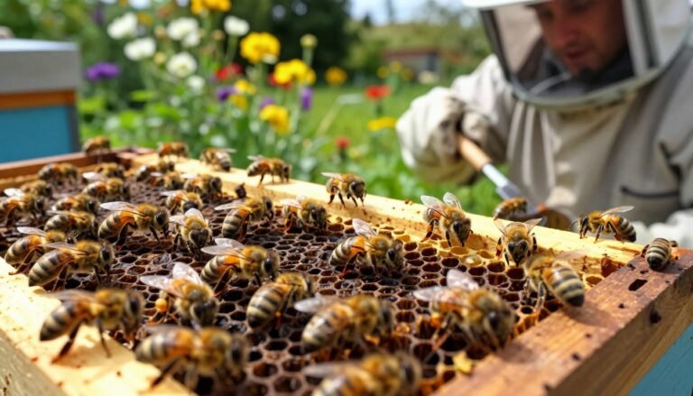
Routine frame-by-frame checks reveal subtle changes in cells before cases escalate. Start each inspection in early spring and again in autumn to compare patterns over time.
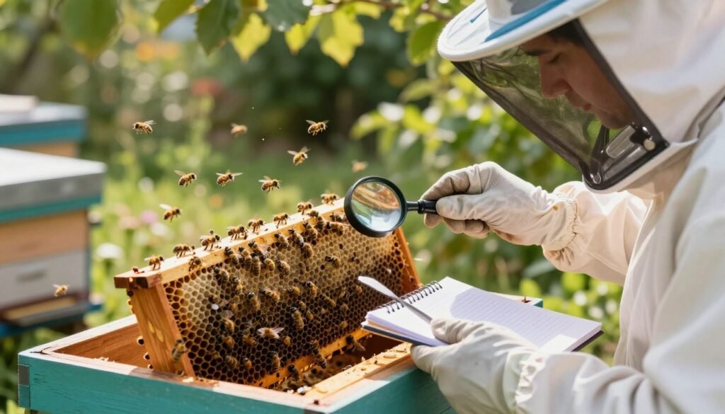
Systematic method: open one hive at a time, loosen top bars, and view frames against light. Note any off-color larva or unusual cap texture and mark frames for follow-up.
Record keeping helps. Photograph suspect areas and log frame numbers, so you can track spread within colonies and across hives.
If you find uncertain signs, submit samples to a lab for confirmation. In Iowa, inspections are provided at no fee — contact the State Apiarist at (515) 725-1481 for help.
When to involve specialists: if multiple frames show odd cells or several hives present similar signs, call for testing or assistance. Familiarity with healthy colonies makes spotting problems faster and reduces accidental spread via shared equipment.
“Early, consistent inspections and lab confirmation when needed are the most reliable defenses against infection.”
- Inspect in early spring and autumn.
- Note unusual caps, off-color larvae, and patterns across frames.
- Use lab testing when unsure and sanitize equipment between hives.
- See practical inspection aids like deadout inspection tips and thermal analysis methods.
Managing and Controlling Infected Colonies
Controlling an infected colony starts with accurate diagnosis and strict handling of frames.
Act quickly. If signs point to foulbrood, isolate the hive and limit movement of equipment and bees. Worker activity such as uncapping diseased cells can increase spore spread across the apiary.
The Role of Antibiotic Treatments
Terramycin is useful only as a preventative measure. It does not destroy spores of american foulbrood and cannot cure an afb infected colony.
Tylosin (Tylan) is an alternative and may help where resistance emerges. Beware: extender patties can under-dose if consumed too slowly. Proper delivery and dosing matter for effective treatment.
“Destruction of bees and comb remains the only reliable elimination method for confirmed american foulbrood.”
| Action | When to use | Key note |
|---|---|---|
| Antibiotic prevention | High risk apiaries | Does not kill spores |
| Tylosin treatment | Resistance concerns | Follow label dosing |
| Burn and replace | Confirmed afb infected cases | Reliable eradication |
| Sanitize equipment | After removal | Reduce cross-colony spread |
For official management guidelines and legal steps, consult disease management resources.
Best Practices for Apiary Sanitation
A sound cleaning plan helps beekeepers decide whether to disinfect gear or replace it.
Methods for Disinfecting Woodenware
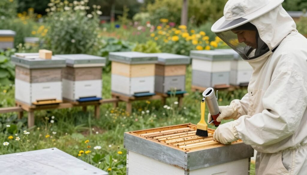
Boiling with lye removes wax and kills many contaminants. Use one can of lye per 15 gallons of water and boil wooden frames for about 20 minutes. Rinse thoroughly with clean water and let combs dry before reuse.
Scorching and torching work well for surface sterilization. Scorch interior faces with a propane torch until blackened to remove propolis and residual honey. This method helps when full replacement is not feasible.
When to Use Fire for Eradication
Destruction by fire remains the most reliable option for confirmed american foulbrood or afb infected equipment. Spores can persist for decades, so burning contaminated combs and frames stops further spread.
Where burning is impractical, consider ethylene oxide fumigation or irradiation services if available. Beekeepers must weigh cost versus risk and follow local regulations for disposal and treatment.
“Safe, timely sanitation and clear choices about equipment reduce the chance of a single infection impacting multiple colonies.”
| Action | Use when | Key note |
|---|---|---|
| Boil in lye water | Contaminated woodenware, reusable frames | 1 can lye / 15 gal; 20 minutes |
| Propane torch | Surface wax/propolis removal | Blacken interior; follow safety rules |
| Burn equipment | Confirmed american foulbrood | Definitive eradication of spores |
| Fumigation / irradiation | When available and legal | Costly but effective alternative |
Conclusion
Consistent, methodical frame reviews let keepers catch trouble while it is still manageable. Regular inspection of the hive is the clearest way to notice early symptoms of diseases like american foulbrood.
Maintaining a healthy colony means vigilance for dead larvae, odd cell appearance, and changes at each stage from larva to pupae. Quick action—sample, test, and isolate—limits spread to other colonies.
Control depends on sound equipment management and prompt treatment or removal when infection is confirmed. For official guidance see the american foulbrood guidance and practical notes on varroa resistance signs.
FAQ
What causes color changes in brood stages?
Multiple agents can alter the appearance of developing young in a comb. Bacterial infections such as American foulbrood and European foulbrood, fungal growths, viral problems, and poor feeding all produce visible shifts. Environmental stress, low pollen availability, and chilling of the comb also create abnormal hues and textures in affected cells.
How can I tell healthy comb patterns from unhealthy ones?
Healthy frames show uniform, plump white larvae, neat cell cappings, and consistent brood distribution. Look for smooth glossy cappings and evenly spaced brood. Patchy areas, sunken caps, or inconsistent development across the frame usually indicate trouble.
What are the key signs of American foulbrood?
American foulbrood causes scattered or sunken caps, darkened remains that may stretch into a ropy thread, and a foul odor in severe cases. Once present, the infection can produce hard scales that cling to the cell base and resist removal, indicating a major contamination requiring strict control.
How does European foulbrood differ in appearance?
European foulbrood often shows twisted or ruffled larvae and uneven brood patterns without the hard, adherent scales of American foulbrood. Dead individuals usually appear yellow to brown and may lack the sticky ropiness seen with the other disease.
Can fungi or viruses produce similar symptoms?
Yes. Fungal organisms often produce fuzzy growth or unusual textures on remains, while some viruses cause irregular development and discoloration without bacterial signs. Lab testing is the only way to confirm the specific pathogen when visual signs overlap.
What diagnostic steps should I take during a hive inspection?
Conduct a systematic frame-by-frame check, note brood pattern and cap condition, and record any abnormal cells. Collect representative samples of suspect material for lab analysis. Use a clean hive tool and avoid moving contaminated frames between colonies.
When is laboratory testing necessary?
If you observe atypical coloration, sunken or perforated caps, ropy remains, or persistent unexplained brood loss, send samples to your state apiary diagnostic lab. Testing confirms the pathogen and guides legal and treatment decisions.
What treatment options exist for infected colonies?
Management depends on diagnosis. For some bacterial diseases, licensed antibiotics under veterinary guidance can suppress infection but may not eliminate spores. In severe or legally regulated cases, burning frames and equipment or complete colony destruction may be required to stop spread.