In 1665, English scientist Robert Hooke published Micrographia, the first major book of microscope observations — and among its sixty detailed plates was the first scientific illustration of a bee’s anatomy ever seen through a lens, a close study of the sting that revealed something no beekeeper working by eye alone had ever documented: the sting itself is barbed. It’s easy to take for granted today that a bee sting has backward-facing barbs, since it’s common knowledge repeated in almost every beginner beekeeping guide — but in 1665, that fact didn’t exist yet. Hooke’s magnified drawing is where it entered recorded science.
Key Takeaways
- Robert Hooke published Micrographia in 1665, at age 30 — the first major scientific work built around microscope observations, and an immediate popular success; Samuel Pepys called it “the most ingenious book that ever I read in my life” in his diary that same January.
- The book’s sixty numbered “Observations” covered a wide range of magnified subjects, from a flea and a louse to the point of a needle — and a dedicated study of a bee’s sting, presented as two detailed engraved figures.
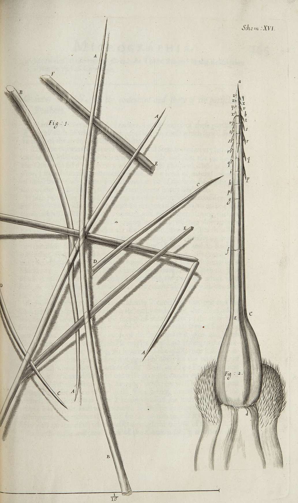
- Hooke’s illustration documented that the bee’s sting carries small backward-pointing barbs, a structural detail invisible to the naked eye and unrecorded before magnified observation made it visible.
- Hooke built his own compound microscope for this work, adding a water-filled lens beside it to focus lamplight onto specimens — an early, practical solution to the lighting problem that limited what earlier microscopists could actually see and draw.
- Micrographia predates the more famous bee-anatomy breakthroughs on this site by decades — Jan Swammerdam’s dissection proving the “king bee” was a queen came roughly a decade later, and François Huber’s observation hive work followed more than a century after that.
Table of Contents
- The Book That Started Microscopy as Popular Science
- What the Bee Sting Plate Actually Showed
- The Microscope Behind the Drawings
- Where This Fits in the History of Bee Science
- Why It Still Matters
- Frequently Asked Questions
The Book That Started Microscopy as Popular Science
Micrographia wasn’t a specialist’s technical manual — it was, by the standards of 1665 London, a genuine bestseller, and the first major book built entirely around what a compound microscope could reveal about ordinary objects. Robert Hooke, then a 30-year-old member of the Royal Society, organized the work as sixty numbered “Observations,” each pairing a large, carefully engraved plate with Hooke’s written description of what the magnification revealed. The range was deliberately broad — the edge of a razor, the point of a needle, a piece of cork (where Hooke coined the biological term “cell,” seeing the honeycomb-like structure of plant tissue for the first time), a flea, a louse, and a fly’s compound eye all received the same close treatment. Samuel Pepys, the naval administrator whose diary remains one of the great firsthand records of the period, stayed up until two in the morning reading it and called it, in his own diary entry for January 21, 1665, “the most ingenious book that ever I read in my life.”
What the Bee Sting Plate Actually Showed
Among Micrographia’s sixty observations, Hooke devoted detailed attention to the sting of a bee, presenting it across two figures on a single engraved plate. The magnified view revealed a level of structural detail no beekeeper handling a live bee could have seen with the naked eye: the sting carries small, backward-angled barbs along its length, arranged in a way Hooke rendered with careful, labeled precision — the same meticulous labeling style he used across the book’s other plates, marking specific structural features with letters referenced in his accompanying text. That barbed structure is exactly why a honey bee’s sting typically remains lodged in flesh after a sting (tearing loose from the bee in the process, which is fatal to a worker honey bee) — a piece of everyday beekeeping knowledge that traces, in recorded scientific history, directly back to this one magnified 17th-century drawing.
The Microscope Behind the Drawings
Hooke didn’t just describe what he saw — he built and refined the instrument that let him see it. He constructed a compound microscope fitted with a new screw-operated focusing mechanism of his own design, solving a practical usability problem earlier microscopists struggled with. Just as important as magnification was illumination: Hooke placed a water-filled lens beside the microscope to focus lamplight onto his specimens, brightening subjects that would otherwise have been too dim and low-contrast to draw accurately at high magnification. That combination — a workable focusing mechanism plus a genuine lighting solution — is a large part of why Micrographia’s plates hold up as genuinely observational rather than speculative; Hooke was drawing what the instrument actually showed him, not filling in gaps with guesswork.
Where This Fits in the History of Bee Science
Micrographia’s 1665 publication date puts it well before most of the bee-science milestones this site has already covered individually. Jan Swammerdam’s dissection proving the “king bee” was actually a queen came roughly a decade later, in the 1670s, using an entirely different method — anatomical dissection under magnification rather than surface observation of an intact structure. François Huber’s observation-hive breakthroughs followed more than a century after that, in the 1790s, and Johann Dzierzon’s discovery of bee parthenogenesis came later still, in the 1840s. Hooke’s bee-sting plate isn’t the deepest of these discoveries in terms of what it revealed about bee biology or behavior — it’s a single anatomical structure, not a colony-level insight — but it’s very plausibly the earliest of the bunch, predating even Swammerdam’s more famous queen-bee dissection, and it marks the moment magnified observation of bee anatomy entered recorded Western science at all.
Why It Still Matters
Micrographia’s bee-sting plate isn’t just a historical curiosity — it’s a clean example of how a single well-documented observation, made with a genuinely new instrument, can outlive the observer by centuries. The barbed structure Hooke drew in 1665 is the same structural detail every modern guide to bee anatomy and sting mechanics still describes, just confirmed since then by far more sophisticated imaging. It’s also a reminder that “ancient” and “modern” bee science aren’t the only two categories worth knowing — a meaningful amount of what’s now common knowledge about bees arrived in a specific, datable moment, from a specific person, using a specific new tool, long before the 19th- and 20th-century breeding and behavior science most beekeeping history content focuses on.
Frequently Asked Questions
What was Micrographia?
Micrographia was a 1665 book by Robert Hooke, the first major work built around microscope observations, presenting sixty detailed engraved plates of magnified subjects including a bee’s sting, a flea, a louse, and the structure of cork.
What did Hooke’s bee sting illustration actually show?
It showed, for the first time in recorded science, that a honey bee’s sting carries small backward-pointing barbs — the structural detail that causes the sting to remain lodged in flesh after use.
Did Hooke build his own microscope?
Yes. He built a compound microscope with a new screw-operated focusing mechanism and added a water-filled lens to focus lamplight onto specimens, solving both the focusing and lighting problems that had limited earlier microscopists.
How does this compare to Jan Swammerdam’s work on bees?
Hooke’s bee-sting observation, from 1665, predates Swammerdam’s dissection proving the “king bee” was a queen by roughly a decade, and used a different method — surface magnification of an intact structure rather than internal dissection.
Was Micrographia popular when it was published?
Yes — it was a genuine popular success for its time. Diarist Samuel Pepys stayed up until 2 a.m. reading it and called it, in his own January 1665 diary entry, “the most ingenious book that ever I read in my life.”
For further reading, see the Royal Society.