This article clears up a common beekeeping misconception and helps you protect your hive and colony health.
Randy Oliver’s May 2019 report in the American Bee Journal challenged the idea that one condition directly causes the other. Many apiarists assume a direct link, but careful research shows correlation does not equal causation.
We will explain how each disease works, why confusion persists, and what testing and management steps can help keep your honey production steady. Proper identification matters for treatment and for avoiding needless interventions that harm the colony.
Thanks for taking the time to read this article. Our goal is to give clear, practical guidance so you can manage your hive with evidence-based practices.
Key Takeaways
- Correlation between the two conditions is common, but causal proof is lacking.
- Microscopy and testing are needed to confirm infection before treatment.
- Environmental stressors often explain concurrent symptoms in a colony.
- Accurate ID preserves honey yields and reduces unnecessary treatments.
- Review Randy Oliver’s analysis for detailed research context: repeat after me: nosema does not cause.
Understanding the Basics of Bee Health
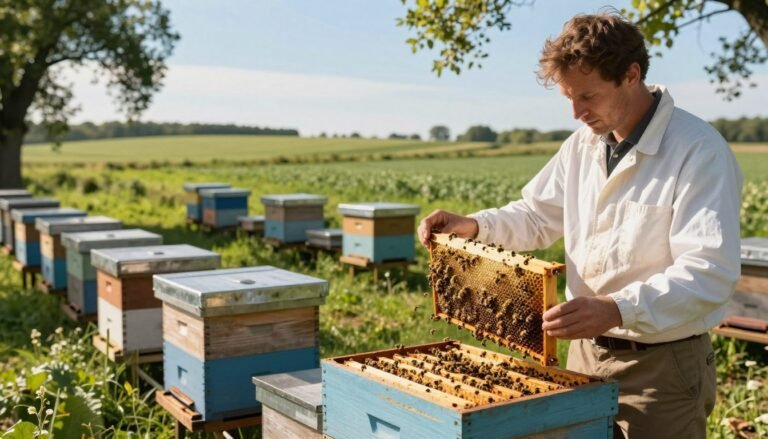
Healthy hives start with daily checks and attention to basic colony needs.
The Importance of Hive Hygiene
Clean equipment and routine cleaning limit disease spread and keep honey stores safe.
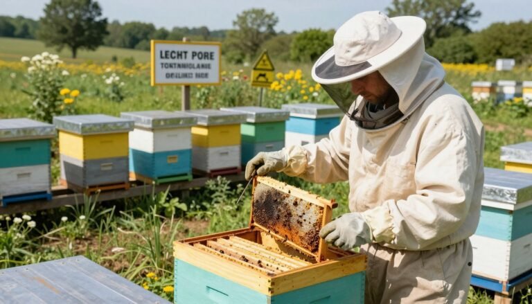
Randy Oliver inspected 40 hives after almond pollination and showed that even strong colonies can face unexpected trouble.
Good ventilation, dry combs, and removed debris reduce stress on the brood and adult worker bee populations.
Recognizing Early Warning Signs
Beekeepers should watch for fecal spotting on the landing board or hive front. That pattern often signals confinement from cold weather.
Spotting can peak each year during early spring, when colonies are most vulnerable after long winter or pollination work.
Quick action matters: distinguish a simple case from a more serious infection to choose the right response.
| Issue | Early Sign | Immediate Action |
|---|---|---|
| Confinement stress | Fecal spotting at entrance | Improve ventilation; clear snow/ice; inspect frames |
| Gut infection | Persistent spotting; weak foragers | Sample for lab testing; consider treatment under guidance |
| Poor hygiene | Debris, mold, or wet combs | Clean hive parts; replace damaged comb; monitor feed |
For more details on identifying early signs, consult resources such as signs of a colony having nosema.
Defining Dysentery in Honey Bee Colonies
Forced confinement during cold weather often leads to visible sanitation problems at the hive entrance.

At its core, dysentery is an excess of water in the gut that affects normal waste control. Honey bees can hold feces for months to avoid soiling combs. When they cannot take cleansing flights, the rectum may distend and fill much of the abdomen.
The presence of yellow or brown streaks on combs or the front of a hive is a major problem needing quick action. These signs point to confinement stress rather than an active infection, though poor sanitation can help pathogens spread.
“A full rectum is a natural physiological state for a bee waiting for a flight; spotting shows the flight window has closed.”
- Key note: This condition is not caused by nosema spores, and it should be treated as a hygiene issue.
- Manage ventilation and allow flights when safe to reduce buildup of water in the gut and lower the risk to the colony.
| Condition | Primary Sign | Immediate Response |
|---|---|---|
| Confinement gut overload | Fecal streaks on combs or hive front | Improve ventilation; clear entrance; monitor flights |
| Sanitation risk | Wet or moldy combs | Replace combs if needed; clean equipment |
| Possible pathogen spread | Persistent spotting; weak foragers | Sample for lab testing; follow treatment guidance |
Exploring the Nature of Nosema Disease
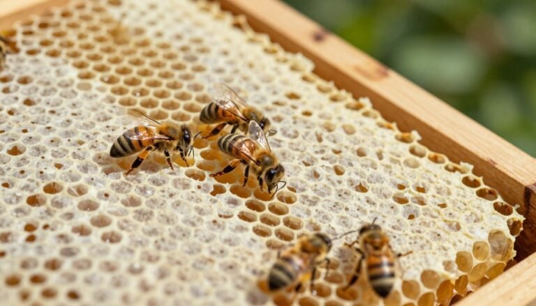
Microsporidian infection targets the gut lining of the honey bee and can quietly reduce colony strength over time.
Nosema Apis vs Nosema Ceranae
Nosema apis historically caused seasonal bouts that often showed white fecal streaking and clearer timing patterns.
Nosema ceranae emerged during the 2000s and behaved differently; it can persist through warm months and sometimes triggers colony collapse.
How Spores Affect the Midgut
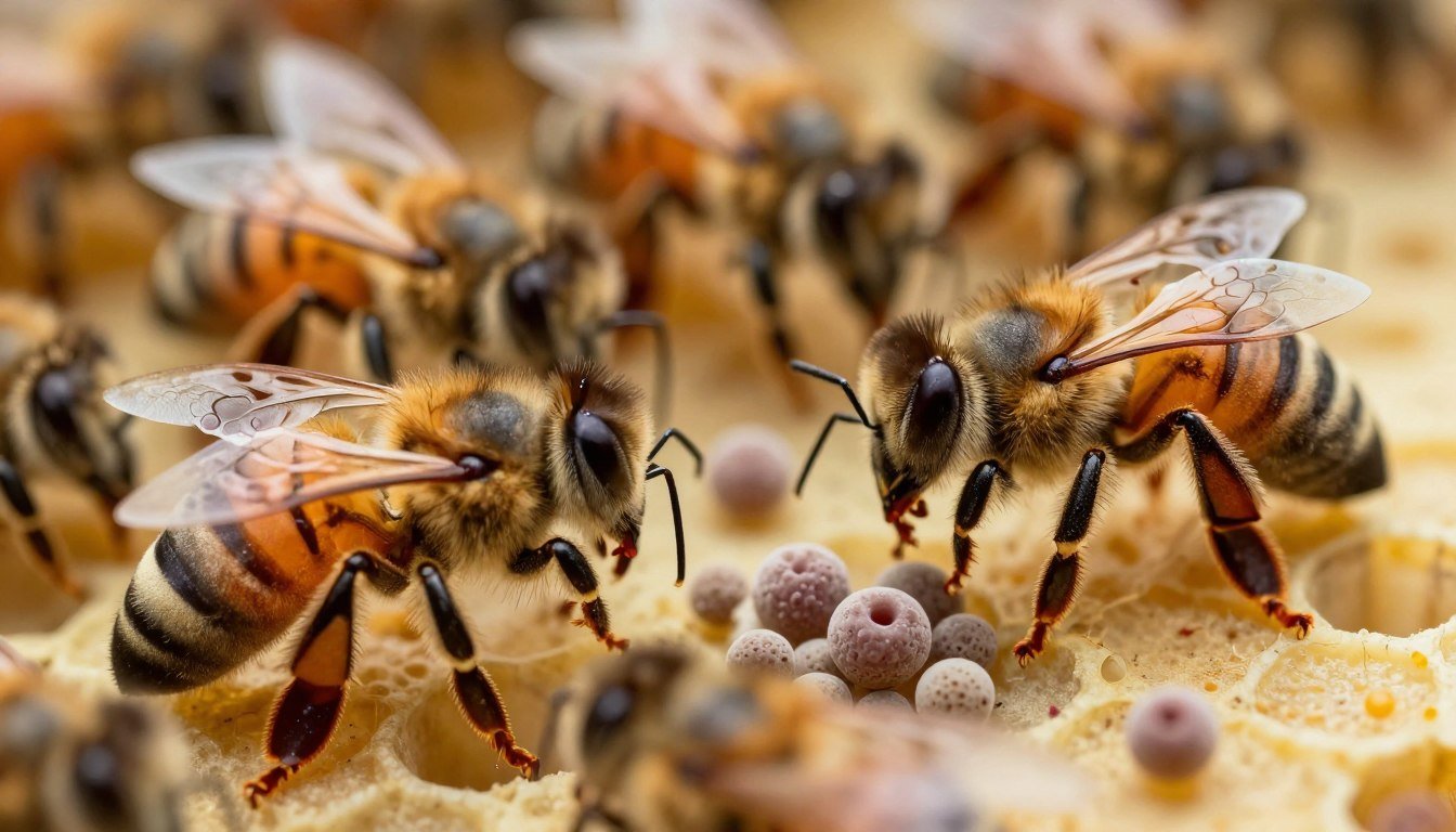
The parasite injects tiny spores into epithelial cells of the midgut. Once inside, the spores multiply and block enzyme production.
This interference causes poor digestion and malnutrition, and severe infection may lead to the death of individual workers.
According to the OIE Terrestrial Manual 2008, nosemosis can deplete a colony, leaving the queen with few workers to maintain brood and honey stores.
“Nosema disease is a microsporidian infection that directly damages midgut cells and undermines colony resilience.”
- Signs often include a dwindling population while combs and sealed brood still look normal.
- Nosema spores reproduce rapidly and change how bees process food.
Investigating the Link Between Dysentery vs Nosema in Bees
When common symptoms appear together in early spring, it can mislead even experienced apiarists about cause and effect.
Extensive research has not produced experimental proof that nosema causes dysentery. Randy Oliver has noted no study that confirms a direct causal chain from spore infection to visible gut overload.
Most published data show a clear correlation during stressful periods. Cold, confinement, or poor forage can produce fecal spotting and reduced colony strength at the same time that infections rise.

“Correlation does not equal causation — separate problems can co-occur without one causing the other.”
Key points to remember:
- Laboratory trials do not support the claim that spores routinely cause cause dysentery.
- If infection were the primary driver, we would expect mass spore transmission and rapid colony collapse, which is not typical.
- Distinguish environmental stress from pathogen-driven illness before treating the hive.
For practical prevention tips and further analysis, see preventing dysentery.
Why the Misconception Persists in Beekeeping
Longstanding beliefs about what causes gut spotting often survive because anecdotes travel faster than studies.
Reports with dramatic photos and short explanations made a simple story easy to repeat. Over time, that story became accepted as fact by many practitioners.
The Dangers of Repeating Unverified Information
Unchecked claims harm management choices. When people assume nosema causes dysentery, they may treat the hive for the wrong issue.
Authors and trainers who cite secondary sources without checking primary research amplify the problem. This reduces trust in valid studies and slows progress.
“Question common wisdom; demand evidence before you change your management plan.”
- Myths lead to wasted treatments and lost honey.
- Confusion makes it harder for beekeepers to diagnose a real issue.
- Good research helps find the true causes and solutions.
| Claim | Evidence Level | Risk if Repeated |
|---|---|---|
| nosema causes dysentery | Low — limited experimental support | Misdirected treatment; resource loss |
| Confinement causes gut spotting | High — strong observational support | Better ventilation and hygiene actions |
| Spore load explains all cases | Moderate — varies by study | Overreliance on medication |
Thanks for reading this article. Stay critical, seek primary research, and protect your hive with informed choices.
Historical Perspectives on Bee Pathogens
Early twentieth-century experiments set the foundation for how we interpret gut infections and hive sanitation today.

G.F. White spent nine years examining nosema apis and published a USDA bulletin in 1919.
His work showed that spores and seasonal stress could coexist, but one did not always cause the other.
Decades later, Dr. Leslie Bailey added practical field notes. In 1967 he reported that dysentery occurred in colonies with and without heavy infection loads.
These early studies challenged simple cause-and-effect stories and urged careful testing before treatment.
Respecting this history helps modern keepers avoid habits based on anecdotes rather than data.
“Long-term observation and controlled trials first pointed away from a direct causal link.”
| Study | Year | Key Finding |
|---|---|---|
| G.F. White, USDA bulletin | 1919 | Nine-year experiments; no direct link between spores and seasonal gut overload |
| Leslie Bailey, field report | 1967 | Observed dysentery across colonies regardless of spore levels |
| Modern reviews | Recent years | Reaffirm early conclusions and call for microscopy and lab testing |
The Role of Environmental Factors in Hive Health
Extended confinement inside the hive forces worker bees to retain waste until they can take cleansing flights.
https://www.youtube.com/watch?v=W_3J1NYbtCc
Cold weather is the main trigger for this stress. When honey bees cannot fly for days or weeks, the rectum fills and spotting appears on combs.
That accumulation of water in the gut is often a natural response to harsh conditions, not necessarily a sign of disease like dysentery or nosema. Treating without testing risks unnecessary intervention.
Practical steps help reduce harm. Monitor local weather closely and check colony ventilation. Ensure the colony has quality stores and access to clean food.
- Open small vents to lower humidity.
- Clear entrances to allow short flights when safe.
- Rotate or supplement feed to maintain strength.
“Manage the environment first; many sanitation issues follow from confinement rather than infection.”
For a seasonal checklist to prepare hives each spring, review this spring beekeeping checklist.
Diagnostic Techniques for Accurate Identification
A clear diagnosis begins with a microscope and a proper sample. Quick field checks are helpful, but lab work confirms what is actually present.

The Necessity of Microscopy
The only reliable way for a beekeeper to confirm a spore-based infection is to view samples under magnification.
Visual signs such as fecal spotting on combs cannot prove an active case. Those marks may reflect confinement stress rather than an active infection.
- Use a well-rated scope, such as the Omano 36, to spot small, elongated ovals that mark nosema spores.
- Crush several dead worker bee abdomens and examine the slurry on a slide for consistent results.
- Research shows experienced keepers often misclassify cases without a microscope.
“Accurate identification is the first step toward the right response for a colony.”
| Step | What to look for | Outcome |
|---|---|---|
| Sample prep | Crushed abdomens on slide | Clear view of spores or absence |
| Microscopy | High magnification, bright field | Confirm whether treatment is needed |
| Follow-up | Record counts and symptoms | Decide on management or environmental fixes |
For a practical guide on lab diagnosis and treatment options, review diagnosing and treating nosema.
Managing Colony Stress and Sanitation
A clean hive entrance and dry combs cut transmission risk and keep colonies resilient.

Proper sanitation is the best defense against pathogens, including nosema spores. Remove heavily soiled frames and replace rotten comb to lower contact with fecal material.
When workers defecate inside the hive due to gut overload, the amount of waste on frames becomes a vector for further infection. That contamination raises transmission rates and stresses the entire colony.
Manage stress by improving ventilation, offering adequate stores, and avoiding unnecessary disturbance during cold snaps. A strong colony resists secondary problems far better than a weakened one.
“At the point where a hive is heavily contaminated, the risk of total colony death becomes very real.”
- Keep entrances clear for short cleansing flights when safe.
- Rotate comb and sanitize tools after heavy soiling events.
- Sample suspect workers and consider lab testing before medicating.
| Risk | Action | Expected Result |
|---|---|---|
| Heavy fecal contamination | Replace frames; deep clean hive | Lower spore contact; reduce spread |
| High colony stress | Improve ventilation; supplement feed | Faster recovery; stronger forager return |
| Confirmed infection | Follow diagnostic guidance; targeted treatment | Controlled outbreak; protect honey stores |
For detailed protocols and research-backed tips, review the nosema and dysentery analysis. Thanks to keepers who prioritize hive hygiene — their work saves colonies.
Implications for Modern Apiary Management
Successful beekeeping now depends on integrating lab diagnostics with sound field practices. Microscopic checks and routine hive care let a beekeeper act on facts, not hearsay.
Modern research shows that Nosema ceranae and Nosema apis behave differently across the year. Track seasonal trends and use microscopy to confirm spores before treating.

Focus on reducing stressors. Weather, scarce forage, and water shortages raise the risk of dysentery and related disease. Strong colonies survive tough springs better when managers provide dry combs and clear entrances for short flights.
“Base management decisions on diagnostics and environmental fixes rather than routine medication.”
- Monitor: periodic spore counts and health checks.
- Maintain: painted, well-vented hive bodies and clean equipment — see maintenance and painting of beehive bodies.
- Minimize stress: steady food and access to water.
| Focus | Action | Result |
|---|---|---|
| Diagnostics | Microscope sampling for spores | Accurate treatment decisions |
| Environment | Improve ventilation and water access | Fewer sanitation issues and stronger colonies |
| Management | Rotate comb; replace soiled frames | Lower contact with infection and better honey quality |
Conclusion
Good management hinges on distinguishing environmental stress from true infection.
There is no scientific evidence that confirms the claim that nosema causes dysentery. Careful review of research shows the two can occur together but one does not necessarily cause the other.
Gut overload often stems from water accumulation and confinement, while nosema is a parasitic problem confirmed only by microscopy. Look for nosema spores and, when relevant, identify strains such as nosema ceranae.
Practical steps—clear the entrance, dry wet comb, and sample suspect workers—protect the hive and its honey stores. Accurate ID keeps treatments targeted and avoids needless harm to the colony.
Thanks for reading this article and for using verified research to guide your apiary care.
FAQ
What causes foul fecal staining inside the hive?
Staining on frames and combs usually comes from digestive upset that forces workers to defecate inside the colony. Cold weather, limited cleansing flights, wet or poor forage, and infections that disrupt the gut can all contribute. Good ventilation, access to clean water, and routine hive checks help reduce buildup and keep combs clean.
Can a microsporidian infection lead to inside-hive soiling?
Yes. Microsporidia like Nosema apis and Nosema ceranae damage the midgut epithelium. Infected workers may lose normal control of bowel movements and soil combs. Lab confirmation is required to link an infection directly to the staining you see.
How do I tell if a colony is suffering from a gut pathogen versus weather-related mess?
Weather problems produce sporadic staining after prolonged cold or rain that limits flights. Pathogen-related staining often appears alongside other signs: weak brood rearing, reduced forager return, scattered dead workers at the hive entrance, and increased spring mortality. Microscopic spore counts or veterinary testing confirm infection.
Are there visible differences between staining caused by disease and normal old honey or propolis?
Yes. Fecal stains are irregular, yellow-brown to dark, and occur on outer comb edges, frames, and covers. Old honey and propolis show more uniform coloration and texture. Regular inspections and documenting changes over time make differentiation easier.
When should I send samples for laboratory diagnosis?
Submit samples when you see persistent staining, increased dead workers, poor spring build-up, or unusual adult mortality. A lab can perform microscopy to detect spores and quantify infection intensity, guiding treatment and management decisions.
What treatments control midgut microsporidia effectively?
Fumagillin has been used historically against Nosema apis, but efficacy varies with species and regulations. Management focuses on hive hygiene, nutrition, splitting strong colonies, and replacing old comb. Consult local extension services or a veterinary diagnostician before applying chemical treatments.
How important are cleansing flights for colony health?
Very important. Regular flights let workers void waste away from the hive. Cold spring weather or prolonged rain increases inside soiling and disease transmission. Providing open sunny apiary sites and minimizing disturbances helps workers take necessary flights.
Does replacing combs reduce the risk of recurring gut infections?
Replacing old and heavily stained combs reduces spore reservoirs and improves brood rearing. Rotate out combs every few years, especially those with repeated staining or darkened cells, to lower pathogen load and chemical residues.
Can good nutrition and water access prevent midgut infections?
Strong nutrition and steady clean water decrease stress and help immune function. Supplemental feeding in dearth periods, diverse forage plantings, and near-hive water reduce colony stress and lower susceptibility to gut infections.
How do I limit spread between colonies in an apiary?
Practice strict sanitation: avoid sharing frames or tools without disinfection, isolate symptomatic colonies, maintain proper spacing to reduce drifting, and inspect frequently. Promptly remove heavily infected equipment and consider requeening weak colonies to restore vigor.
Are dead bees on the bottom board always a sign of infection?
Not always. Seasonal mortality, pesticide exposure, and predation can leave corpses. A cluster of dead workers with fecal staining or a pattern of increased loss during spring suggests a health issue and warrants further investigation and testing.
Should I change winter management practices to reduce spring problems?
Yes. Avoid starving colonies, ensure good ventilation to reduce moisture, and minimize disturbances that prevent cleansing flights in mild winter days. Early spring feeding and timely inspections help colonies resume normal foraging and sanitation behaviors.